Computing mit Gehirnorganoiden für die Entscheidungsfindung in der Robotik
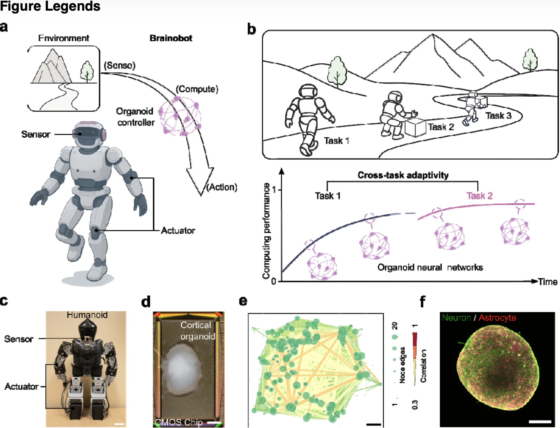

a. Schematic diagram of the Brainobot framework for controlling a humanoid. The organoid controller receives sensory inputs from the humanoid, processes the information, and outputs action commands that drive corresponding movements through organoid neural networks. b. Schematic illustration of Brainobot performing real-world tasks and the hypothesized corresponding learning curves over time and across different tasks through reshaping of organoid neural networks. c. Image of a humanoid implemented with remote control of an organoid controller. d. Physical setup of an organoid controller by mounting a cortical organoid onto a MaxOne microelectrode array (MEA) chip, scale bar = 500μm. e. Representative functional connectivity map of a 3-month-old cortical organoid on MEA, scale bar = 200μm. Edge color and width indicate the degree of correlation between neuron units, while node size reflects the number of connections per neuron. f. Whole-mount immunostaining of a 3-month-old cortical organoid showing complex three-dimensional neuronal networks with mature neurons (MAP2+) and astrocytes (GFAP+), scale bar = 200μm.
a. Schematische Darstellung des Brainobot-Frameworks zur Steuerung eines Humanoiden. Der Organoid-Controller empfängt sensorische Eingaben vom Humanoiden, verarbeitet die Informationen und gibt Aktionsbefehle aus, die über Organoid-Neuralnetzwerke die entsprechenden Bewegungen steuern. b. Schematische Darstellung von Brainobot bei der Ausführung realer Aufgaben sowie der hypothetischen entsprechenden Lernkurven im Zeitverlauf und über verschiedene Aufgaben hinweg durch Umgestaltung der Organoid-Neuralnetzwerke. c. Abbildung eines Humanoiden, der mittels Fernsteuerung durch einen Organoid-Controller gesteuert wird. d. Physikalischer Aufbau eines Organoid-Controllers durch Montage eines kortikalen Organoids auf einem MaxOne-Mikroelektrodenarray (MEA)-Chip, Massstabsbalken = 500 μm. e. Repräsentative Karte der funktionellen Konnektivität eines 3 Monate alten kortikalen Organoids auf einem MEA, Massstabsbalken = 200 μm. Die Farbe und Breite der Kanten zeigen den Grad der Korrelation zwischen den Neuroneneinheiten an, während die Knotengrösse die Anzahl der Verbindungen pro Neuron widerspiegelt. f. Ganzpräparat-Immunfärbung eines 3 Monate alten kortikalen Organoids, die komplexe dreidimensionale neuronale Netzwerke mit reifen Neuronen (MAP2+) und Astrozyten (GFAP+) zeigt, Massstab = 200 μm.
Die, äh, “Studie” gibt's hier (Sicherungskopie).